発明の背景
本発明は、新規アポトーシス誘導剤、カスペースカスケード活性化剤及び制癌剤に関する。
アポトーシスの概念が最初に提唱されたのはKerr、Wylli.Currieによる1972年の報告である(Kerr J.F.R.,Wyllie A.H.and Currie A.R.,Br.J.Cancer,26,239〜257(1972))。アポトーシスは、細胞の縮小、クロマチンの凝縮、DNAのヌクレオソーム単位(約180bp)での断片化などの著しい形態変化によって特徴付けられ、ネクローシス(壊死)が外部刺激の物理的作用による突発的な細胞死であるのに対し、個体維持のために遺伝子に制御された細胞死として定義される。
アポトーシスのメカニズムは、誘導、決定、実行の3つの経路を経ると考えられている。アポトーシスのメカニズムにおいて主要な役割を果たしている決定過程は2つの伝達経路を有している。その1つは、ミトコンドリアを介する経路でありBcl−2ファミリーがこの経路を制御している。もう1つは、カスペースによる自己分解および、カスペースカスケードの下位のカスペースの限定分解による伝達経路、いわゆるカスペースカスケードである。
このアポトーシスの分子メカニズムは、Horvitz、Yuanらが線虫を用いて調べた。彼らは線虫を構成する細胞のアポトーシスに関与する合計14の遺伝子を発見し、アポトーシス実行因子としてced−3、ced−4、抑制因子としてced−9が特に重要であることを見出した。遺伝子同定の結果、ced−3がヒトIL−1β変換酵素(ICE)というシスティンプロテアーゼと相同性が高いことが判明した(Yuan J.,Shaham S.,Ledoux S.,Ellis H.M.and Horvitz H.R.,Cell,75,641〜652(1993))。さらに、ICEの過剰発現によりアポトーシスが誘導されることが確認されたことより、ICEは、ヒトにおいてもアポトーシス実行因子として働いているのではないかと考えられるようになった。しかし、その後報告されたICEノックアウトマウスでは、アポトーシスの機能に殆ど影響はなかった。また、その後、アポトーシスに特徴的なポリ(ADP−リボース)ポリメラーゼの分解が、ICEでは起こらないにもかかわらず、ICEに似たシスティンプロテアーゼによって起こることも明かにされた。これらより、ヒトにおけるアポトーシス実行因子はICEそのものではなく、ICE様の別の酵素ではないかと推測され、ICE以外のICE様酵素がヒトに存在するものと考えられた。
以上から、ICE様酵素の同定がYuanらの報告以降試みられ、ICEを含めヒトには10種類程度のICE様酵素があることがわかった。同定されたICE様酵素は、Asp特異的システィンプロテアーゼということで、酵素名が「カスペース(caspase)」に統一され、報告順にカスペース−1(ICE)〜カスペース−10など番号を付け命名され、一般にカスペースファミリープロテアーゼと呼称されるようになった。
現在、カスペースは基質特異性から大きく3つに分類され、カスペース−1を中心とするI群はインターロイキンの生成と分泌に関与していると考えられ、カスペース−3を中心とするII群はアポトーシスの実行分子の中心と考えられ、カスペース−8/−9に代表されるIII群はカスペースのタンパク質分解カスケードの上流に位置し、アポトーシスのための細胞死シグナルを伝える働きがあると考えられている。
カスペースファミリーは共通の活性中心シークエンスにより、Asp(D)のC末端側を切断する活性を持ち、それぞれ若干異なる基質特異性を持つ。一般にカスペースは、不活性の前駆体として作られ、活性化酵素により2つのサブユニットに切断され、活性型カスペースとなるが、前駆体の活性化における切断部位は、カスペース分子内のAsp残基のC末端側であり、カスペースの基質認識部位と一致している。従って、カスペース自身が別のカスペースを活性化するメカニズムが働いていると推測される。Fasにより誘導されるアポトーシスでは、カスペース−1の活性化の後、カスペース−3様の活性が増大するという現象が見られ、カスペース−3がカスペース−1の下流域に位置することが知られている(Enari M.,Talanian R.V.,Wong W.W.and Nagata S.,Nature,380,723〜726(1996))。このことから、アポトーシスの実行過程では、いくつかのカスペースが連続的に活性化される、いわゆるカスペースカスケードを形成していることが推定される。また、カスペースは、細胞内のタンパク質を分解することにより、アポトーシスの実行過程においても重要な働きをしている。
このように、ヒトで多数のカスペースが存在するのは興味深く、個体を維持するため、複雑なカスペースネットワークが形成されていると考えられている。
しかし、実際にはどのようなメカニズムでカスペースカスケードが活性化されるか、現在のところ未解明の部分が多い。
上述したように、生体内で重要な役割を果たすアポトーシス及びカスペースカスケードは、医薬品の開発においても重要であり、アポトーシス及びカスペースカスケードのメカニズムを利用することにより、アポトーシス誘導のメカニズムが正常に機能しない癌など、細胞増殖性疾患の治療薬を開発することが考えられている。
例えば、特開2000−143532号公報には、薬理活性成分として環状RGD配列を持つ薬剤により、カスペースカスケードを活性化し、アポトーシスを誘導することで細胞増殖性疾患を治療することが示されている。しかしながら、ジミリストイルホスファチジルコリンを有効成分とするようなアポトーシス誘導剤及びカスペースカスケード活性化剤については、これまでに知られていない。
一方、脂質成分を用いた制癌剤は、幾つか知られている。一般に脂質成分を用いた制癌剤は、リポソームや乳剤などの形態で、薬物を運ぶ担体として脂質成分を用いることが殆どであり、脂質成分そのものが制癌剤として作用するとしたものは少ない。脂質成分の癌に対する有効性を示した公報を以下に列挙する。特開平6−256181号公報には、トコフェロールを有効成分とする細胞分化誘導剤が示され、癌の治療・改善剤として有効であることが記載されている。特開平1−203330号公報には、ドコサヘキサエノイル−リゾホスファチジルコリンを有効成分とする制癌剤が記載されている。特開平1−203322号公報にはドコサヘキサエノイルモノグリセリドを有効成分とする制癌剤が記載されている。特開平1−203331号公報には1,2−ジドコサヘキサエノイル−ホスファチジルコリンを有効成分とする制癌剤が記載されている。特開平3−163031号公報にはリン脂質とミセル界面活性剤によって形成した混合分子集合体に抗癌剤を加えた液体膜制癌剤が記載されている。特開平2−11516号公報にはリン脂質の一種を有効成分とした制癌剤が記載されている。既知のリン脂質を利用した制癌剤について、その制癌メカニズムについてはまったく解明されておらず、ましてや制癌活性がアポトーシスによるものであるとしたような研究は知られていなかった。
発明の開示
本発明の第一の目的は、ジ(アルキルカルボニル)ホスファチジルコリン等を有効成分として含有する新規アポトーシス誘導剤を提供することにある。
本発明の第二の目的は、ジ(アルキルカルボニル)ホスファチジルコリン等を有効成分として含有する新規カスペースカスケード活性化剤を提供することにある。
本発明の第三の目的は、ジ(アルキルカルボニル)ホスファチジルコリン等を有効成分として利用する副作用のない制癌剤を提供することにある。
本発明者らは、リン脂質の一種として良く知られるジミリストイルホスファチジルコリン等のグリセロリン脂質に注目し、意外にもこれらの化合物がアポトーシス誘導剤、及びカスペースカスケード活性化剤として作用することを見出し、これが従来の制癌剤と比較して、副作用が無く安全性が高い制癌剤として利用できるという新たな知見を得て本発明を完成した。
即ち、本発明は、有効成分として、ジ(アルキルカルボニル)ホスファチジルコリン、ジ(アルキルカルボニル)ホスファチジルアルカノールアミン、ジ(アルキルカルボニル)ホスファチジルイノシトール、ジ(アルキルカルボニル)ホスファチジルセリンからなる群から選ばれる少なくとも一種を含有することを特徴とするアポトーシス誘導剤を提供する。
本発明はまた、有効成分として、ジ(アルキルカルボニル)ホスファチジルコリン、ジ(アルキルカルボニル)ホスファチジルアルカノールアミン、ジ(アルキルカルボニル)ホスファチジルイノシトール、ジ(アルキルカルボニル)ホスファチジルセリンからなる群から選ばれる少なくとも一種を含有することを特徴とするカスペースカスケード活性化剤を提供する。
本発明また、これらのアポトーシス誘導剤又はカスペースカスケード活性化剤により腫瘍細胞をアポトーシスさせることを特徴とする制癌剤を提供する。
本発明また、ジ(アルキルカルボニル)ホスファチジルコリン等を有効成分として含有してなる分子集合体により腫瘍細胞のカスペースカスケードを活性化させることにより腫瘍細胞をアポトーシスさせることを特徴とする制癌剤を提供する。
本発明はさらに、ジミリストイルホスファチジルコリンと医薬的に許される担体及び/又はアジュバントからなる制癌剤を提供する。
発明を実施するための最良の形態
本明細書において、「アポトーシスさせる」とは、細胞の縮小、クロマチンの凝縮、DNAのヌクレオソーム単位(約180bp)での断片化などの著しい形態変化によって特徴付けられた、医学および生物学において形態学的、生化学的に定義された変化を細胞に生じせしめることであり、ネクローシス(壊死)と異なったプログラム細胞死に至らせることをいうものである。細胞がアポトーシスしているかどうかは、電気泳動によるDNAラダーの観測や、光学顕微鏡による形態観察など、公知の方法によって確認することができる。
また、本明細書において、「カスペースカスケードを活性化させる」とは、カスペースと呼ばれるAsp特異的システィンプロテアーゼ(アスパラギン酸のC末端を加水分解する酵素)により細胞内に構築されるアポトーシスのシグナル伝達ネットワークのなかで、いずれかのカスペースを活性化された状態に至らしめ、カスペースカスケードを活性化させることをいうものであり、必ずしも細胞内の全ての種類のカスペースを活性化させることをいうのではない。また、多様なカスペースのうち、いずれが活性化するかは、細胞の種類や条件などにより異なる場合がある。
カスペースの活性化は、カスペース阻害剤を用いたフローサイトメーターでの阻害実験や、ウエスタンブロット法によるカスペース分解物の検出などの公知の方法を用いることにより推定・確認することができる。
また、本明細書において「腫瘍細胞」とは、いわゆる癌などを構成する細胞、またはそれに由来・関連する細胞であって、一般に腫瘍細胞といわれる細胞であるなら特に限定されない。また、疾患として器官組織内にある細胞だけでなく、ホモジネートした組織から分離したり、また試験用細胞として一般に供される培養細胞や、これら腫瘍細胞由来のハイブリドーマなども含み、ヒト細胞だけに限定されるわけでもない。このような腫瘍細胞としては、例えば、急性白血病、慢性白血病、悪性リンパ腫、多発性骨髄腫、マクログロブリン血症などの造血器腫瘍や、脳腫瘍、頭頸部癌、乳癌、肺癌、食道癌、胃癌、大腸癌、肝癌、胆嚢・胆管癌、膵癌、膵島細胞癌、腎細胞癌、副腎皮質癌、膀胱癌、前立腺癌、睾丸腫瘍、卵巣癌、子宮癌、絨毛癌、甲状腺癌、悪性カルチノイド腫瘍、皮膚癌、悪性黒色腫、骨肉腫、軟部組織肉腫、神経芽細胞腫、ウィルムス腫瘍、胎児性横紋筋肉腫、網膜芽細胞腫などの固形腫瘍を形成する細胞があげられる。
本明細書において、「医薬上許される担体及び/又はアジュバント」という用語は、ジミリストイルホスファチジルコリンと一緒に患者に投与することができ、ジミリストイルホスファチジルコリンの薬理学的活性を損なわない無毒性の担体又はアジュバントを表す。
まず、本発明の第一の態様について説明する。
本発明の第一の態様は、有効成分として、ジ(アルキルカルボニル)ホスファチジルコリン、ジ(アルキルカルボニル)ホスファチジルアルカノールアミン、ジ(アルキルカルボニル)ホスファチジルイノシトール、ジ(アルキルカルボニル)ホスファチジルセリンからなる群から選ばれる少なくとも一種を含有するアポトーシス誘導剤に関する。
本発明のアポトーシス誘導剤は、正常なアポトーシスの機能が抑制されていることによって生ずる種々の疾患の予防又は治療に有効である。このような疾患としては、細胞増殖性疾患、奇形、自己免疫疾患等があげられる。特に、アポトーシスのメカニズムが直接的または間接的に関与している癌などを構成する腫瘍細胞をアポトーシスさせることにより、癌などの細胞増殖性疾患の予防または治療に対して有効である。
従って、本発明はまた、ジ(アルキルカルボニル)ホスファチジルコリン、ジ(アルキルカルボニル)ホスファチジルアルカノールアミン、ジ(アルキルカルボニル)ホスファチジルイノシトール、ジ(アルキルカルボニル)ホスファチジルセリンからなる群から選ばれる化合物を有効成分とする、細胞増殖性疾患、奇形又は自己免疫疾患のいずれかに対する予防剤又は治療剤に関する。
本発明はまた、アポトーシス誘導剤を製造するためのジ(アルキルカルボニル)ホスファチジルコリン、ジ(アルキルカルボニル)ホスファチジルアルカノールアミン、ジ(アルキルカルボニル)ホスファチジルイノシトール、ジ(アルキルカルボニル)ホスファチジルセリンからなる群から選ばれる化合物の使用に関する。
本発明はまた、有効成分として、ジ(アルキルカルボニル)ホスファチジルコリン、ジ(アルキルカルボニル)ホスファチジルアルカノールアミン、ジ(アルキルカルボニル)ホスファチジルイノシトール、ジ(アルキルカルボニル)ホスファチジルセリンからなる群から選ばれる化合物を腫瘍細胞に接触させて腫瘍細胞にアポトーシスを誘導する方法に関する。
本発明はまた、有効成分として、ジ(アルキルカルボニル)ホスファチジルコリン、ジ(アルキルカルボニル)ホスファチジルアルカノールアミン、ジ(アルキルカルボニル)ホスファチジルイノシトール、ジ(アルキルカルボニル)ホスファチジルセリンからなる群から選ばれる化合物を、細胞増殖性疾患、奇形又は自己免疫疾患のいずれかの疾病の患者に投与することを含む、これらの疾病の予防又は治療方法に関する。
本発明で用いる有効成分に含まれる2つのアルキルカルボニルは、同じでも異なっていても良いが、同じであるのが好ましい。アルキルカルボニル基の炭素数は10〜20であるのが好ましく、10〜18であるのがより好ましく、さらに好ましくは12〜16、特に好ましくは14である。また、アルキルカルボニル基におけるアルキル基部分は直鎖でも分岐鎖でもよい。
本発明で用いる有効成分がジ(アルキルカルボニル)ホスファチジルアルカノールアミンの場合、アルカノール部分の炭素数は1〜6であるのが好ましく、1〜3がより好ましく、特に2であるのが好ましい。
本発明で用いる好ましい有効成分は、以下の一般式(1)で表される。
一般式(1)において、R1は、CH3(CH2)m−2である。ここで、mは、10〜18、好ましくは12〜16、より好ましくは14の整数である。
R2は、OCH2CH2N+(CH3)3、OCH2CH2NH2、OCH2CH(NH2)COOH、又は下記式(2)
で表される基である。
本発明で用いる有効成分としては、ジ(アルキルカルボニル)ホスファチジルコリンが好ましい。
一般式(1)で表される化合物として最も好ましいのは、下記一般式(3)で表されるジミリストイルホスファチジルコリン(DMPC)である。
なお、ジミリストイルホスファチジルコリンは、ホスファチジルコリン(phosphatidylcholine)又はレシチン(lecithin)と呼ばれるリン脂質の中の、更にその一種類であり、正式には1,2−ジミリストイル−sn−グリセロ−3−ホスホコリンと呼ばれる。ジミリストイルホスファチジルコリンは、ジミリストイルレシチンやL−α−ジミリストイルホスファチジルコリンと称されることもある。
本発明で用いる有効成分は、単独又は二種以上を混合して使用することができる。二種以上を混合して使用する場合、ジ(アルキルカルボニル)ホスファチジルコリン、特に、DMPCと他の有効成分とを併用するのが好ましい。
本発明で用いられるジ(アルキルカルボニル)ホスファチジルコリン等の有効成分は、天然物由来であっても化学合成品由来であってもよい。天然物由来である場合、動物、植物、微生物などの原料、例えば脳、肝臓、卵黄、大豆、酵母などから公知の方法により抽出・分離・精製して得ることが出来る。また市販のレシチンを原料として、脂肪酸鎖長の違いを利用した公知の分離方法を利用することによりこれらの有効成分を得ることもできる。化学合成品由来の場合、公知の合成化学の知識を駆使することにより、容易にこれらの有効成分を合成することができるが、例えばジミリストイルホスファチジルコリンの場合、グリセロホスフォコリンにミリスチン酸を公知の方法で導入するなどの手法により得ることも出来る。また、市販品を利用してもよく、医薬品グレードの高純度のものが特に好ましい。例えば、DMPCとして、日本油脂株式会社(NOF Corporation)からコートソームMC−4040(COATSOME MC−4040)の商品名で市販されている。
本発明のアポトーシス誘導剤は、有効成分をそれ自体公知の薬理的に許容される担体、賦形剤、崩壊剤、矯正剤、増量剤、希釈剤、溶解補助剤などと混合し、公知の方法に従って医薬組成物、例えば錠剤、カプセル剤、顆粒剤、散剤、粉末剤、丸剤、溶剤、ドリンク剤、注射剤、点滴剤、座剤などの形態に製剤化することができる。このような製剤は経口的もしくは非経口的に投与することができる。
本発明のアポトーシス誘導剤を疾患の治療等に使用する場合の投与量は、投与対象、投与経路、症状などによっても異なるが、血中に投与する場合、一回量として有効成分を1×10⁻¹〜1×10⁻⁶Mの濃度で含む溶液0.01〜100ml/kg体重を1日1〜3回程度投与するのが好適である。有効成分を5×10−2〜5×10−4Mの濃度で含む溶液1〜50ml/kg体重を1日1〜3回程度投与するのがより好ましい。
次に、本発明の第二の態様について説明する。
本発明の第二の態様は、有効成分としてジ(アルキルカルボニル)ホスファチジルコリン、ジ(アルキルカルボニル)ホスファチジルアルカノールアミン、ジ(アルキルカルボニル)ホスファチジルイノシトール、ジ(アルキルカルボニル)ホスファチジルセリンからなる群から選ばれる少なくとも一種を含有するカスペースカスケード活性化剤に関する。
本発明のカスペースカスケード活性化剤は、正常なカスペースカスケード活性化の機能が抑制されていることによって生ずる種々の疾患の予防または治療に有効である。このような疾患としては、細胞増殖性疾患、奇形、自己免疫疾患等があげられる。特に、カスペースカスケードを活性化させることにより、アポトーシスのメカニズムの正常化を促し、癌などを構成する腫瘍細胞等の細胞増殖性疾患の予防または治療に対して有効である。
従って、本発明はまた、カスペースカスケード活性化剤を製造するためのジ(アルキルカルボニル)ホスファチジルコリン、ジ(アルキルカルボニル)ホスファチジルアルカノールアミン、ジ(アルキルカルボニル)ホスファチジルイノシトール、ジ(アルキルカルボニル)ホスファチジルセリンからなる群から選ばれる化合物の使用に関する。
本発明はまた、有効成分として、ジ(アルキルカルボニル)ホスファチジルコリン、ジ(アルキルカルボニル)ホスファチジルアルカノールアミン、ジ(アルキルカルボニル)ホスファチジルイノシトール、ジ(アルキルカルボニル)ホスファチジルセリンからなる群から選ばれる化合物を腫瘍細胞に接触させて腫瘍細胞のカスペースカスケードを活性化する方法に関する。
本発明のカスケード活性化剤として用いられる有効成分としては、アポトーシス誘導剤に関して記載したのと同じである。
本発明のカスペースカスケード活性化剤は、有効成分をそれ自体公知の薬理的に許容される、担体、賦形剤、崩壊剤、矯正剤、増量剤、希釈剤、溶解補助剤などと混合し、公知の方法に従って医薬組成物、例えば錠剤、カプセル剤、顆粒剤、散剤、粉末剤、丸剤、溶剤、ドリンク剤、注射剤、点滴剤、座剤などの形態に製剤化することができる。このような製剤は経口的もしくは非経口的に投与することができる。
本発明のカスペースカスケード活性化剤を疾患の治療等に使用する場合の投与量は、投与対象、投与経路、症状などによっても異なるが、注射剤または点滴剤として投与する場合、一回量として有効成分を1×10⁻¹〜1×10⁻⁶Mの濃度で含む溶液0.01〜100ml/kg体重を1日1〜3回程度投与するのが好ましい。有効成分を5×10−2〜5×10−4Mの濃度で含む溶液1〜50ml/kg体重を1日1〜3回程度投与するのがより好ましい。
最後に、本発明の第三の態様について説明する。
本発明の第三の態様は、有効成分としてジ(アルキルカルボニル)ホスファチジルコリン、ジ(アルキルカルボニル)ホスファチジルアルカノールアミン、ジ(アルキルカルボニル)ホスファチジルイノシトール、ジ(アルキルカルボニル)ホスファチジルセリンからなる群から選ばれる少なくとも一種を利用する制癌剤に関する。本発明の制癌剤として用いられる有効成分としては、アポトーシス誘導剤に関して記載したのと同じである。
本発明の制癌剤は、正常なカスペースカスケードの活性化の機能が抑制されている腫瘍細胞等に有効に作用することによって、そのカスペースカスケードを活性化させ、該腫瘍細胞等をアポトーシスさせることにより癌を予防または治療することができる。本発明の制癌剤の対象疾患は、いわゆる癌であるならばこれらに限定されないが、上記、造血器腫瘍や固形腫瘍に分類される癌、特に急性白血病、慢性白血病、悪性黒色腫、肺癌、肝臓癌、胃癌、悪性脳腫瘍の予防または治療に対して本発明の制癌剤は特に優れている。
従って、本発明はまた、ジ(アルキルカルボニル)ホスファチジルコリン、ジ(アルキルカルボニル)ホスファチジルアルカノールアミン、ジ(アルキルカルボニル)ホスファチジルイノシトール、ジ(アルキルカルボニル)ホスファチジルセリンからなる群から選ばれる化合物を有効成分とする、急性白血病、慢性白血病、悪性黒色腫、肺癌、肝臓癌、胃癌、悪性脳腫瘍の予防剤又は治療剤を提供する。
本発明はまた、制癌剤を製造するためのジ(アルキルカルボニル)ホスファチジルコリン、ジ(アルキルカルボニル)ホスファチジルアルカノールアミン、ジ(アルキルカルボニル)ホスファチジルイノシトール、ジ(アルキルカルボニル)ホスファチジルセリンからなる群から選ばれる化合物の使用に関する。
本発明はまた、有効成分として、ジ(アルキルカルボニル)ホスファチジルコリン、ジ(アルキルカルボニル)ホスファチジルアルカノールアミン、ジ(アルキルカルボニル)ホスファチジルイノシトール、ジ(アルキルカルボニル)ホスファチジルセリンからなる群から選ばれる化合物を、急性白血病、慢性白血病、悪性黒色腫、肺癌、肝臓癌、胃癌、悪性脳腫瘍のいずれかの疾病の患者に投与することを含む、これらの疾病の予防又は治療方法に関する。
本発明の制癌剤は、ジ(アルキルカルボニル)ホスファチジルコリン、ジ(アルキルカルボニル)ホスファチジルアルカノールアミン、ジ(アルキルカルボニル)ホスファチジルイノシトール、ジ(アルキルカルボニル)ホスファチジルセリンからなる群から選ばれる少なくとも一種を、それ自体公知の薬理的に許容される担体、賦形剤、崩壊剤、矯正剤、増量剤、希釈剤、溶解補助剤などと混合し、公知の方法に従って医薬組成物、例えば錠剤、カプセル剤、顆粒剤、散剤、粉末剤、丸剤、溶剤、座剤などの形態に自由に製剤化することができる。このような製剤は経口的もしくは非経口的に投与することができる。特に、ドリンク剤、注射剤または点滴剤として用いるのが好ましい。
注射剤または点滴剤、ドリンク剤として投与するとき、ジ(アルキルカルボニル)ホスファチジルコリン等の有効成分を分子集合体に含有させて投与せしめることが好ましく行われる。これらの有効成分を含有させる分子集合体の例としては、脂肪乳剤、高分子ミセル、アルブミンなどのタンパクとの複合体、リポソームなどが挙げられるが、有効成分が水性溶媒中でそれ自身、リポソームを形成する機能を持つなどの理由から、リポソームが最も好ましく挙げられる。
リポソームを形成する場合、有効成分濃度を5×10⁻⁴〜1.0×10⁻¹Mとするのが好ましく、1×10⁻²〜5×10⁻²Mとするのがより好ましい。
本発明におけるリポソームは、公知のいかなる方法によって形成してもよい。例えば薄膜法、逆相蒸発法、凍結融解法、エタノール注入法、高圧乳化法、超音波分散法、透析法、エクストルージョン法など、公知の製造方法を適宜利用することができ、更には特開平9−87168号公報に記載される方法などを利用してもよい。
リポソーム調製に使用できる水性溶媒としては、単なる水を用いても良いし、少量のエタノールなどを含む水性混合溶媒であってもよく、また血漿などであってもよいが、好ましくは局方注射用水や蒸留水、超純水などが用いられる。水性溶媒に公知の生理活性物質や、蛋白質、緩衝物質や各種の塩、血漿、糖などを溶解させ、溶液状としたものを用いてもよい。
このようにして得られるリポソームは、限外濾過、遠心分離、ゲル濾過等の方法によって精製してよく、また濃縮や希釈等の操作を自由に行ってもよい。
また、本発明におけるリポソームには、本発明の目的に反しない程度に、公知のリポソーム構成成分を含有させて用いてもよい。具体的には、卵黄レシチン、大豆レシチン、水添卵黄レシチン、水添大豆レシチン、ホスファチジルグリセロールなどのリン脂質が挙げられる。
またリポソームの膜安定成分として少量のコレステロールを用いることもできるが、多量のコレステロールは、ジミリストイルホスファチジルコリンの制癌活性を減ずる恐れがあることから好ましくない。また、荷電物質としてホスファチジン酸や脂肪酸を加えても良い。
さらに、分散安定剤としてポリオキシエチレンドデシルエーテルに代表される、炭素数10〜20のアルキルPEGやトライトン、プルロニックなどの商品名で知られる界面活性剤を加えることができる。このうち炭素数12〜14のアルキルPEGが好ましい。特に、ジ(アルキルカルボニル)ホスファチジルコリンとミセルを形成する界面活性剤とから形成されるハイブリッドリポソームを用いるのが好ましい。中でも、DMPCと炭素数10〜16、EO数2〜30のアルキルPEG(好ましくは炭素数12〜14、EO数2〜24)とを組み合わせるのが好ましい。
また、本発明において、糖をリポソームの分散安定化に寄与し制癌活性を高める目的で水性溶媒中に特に好ましく添加することができる。利用できる糖としては単糖、二糖、オリゴ糖、多糖など公知の糖を目的に応じて特に制限されることなく自由に使用することができるが、特に、蔗糖やトレハロースなどが好ましい。またその添加量も適宜選択されて良い。また、酸化防止剤としてα−トコフェロールなどをリポソームに加えても良い。
調製されるリポソームの直径は、医学的に許容される範囲内であればいかなる直径であってもよいが、20nm〜1μm程度が好ましく、より好ましくは30nm〜300nm程度、さらに好ましくは制癌活性を高めるなどの理由から40nm〜180nm程度がよい。リポソームは、水性溶媒中に均一に分散するよう調製されるのがよく、沈殿、凝集などが見られる場合、主に物理的な理由から制癌剤として用いるのが困難になるので好ましくない。
本発明の制癌剤を疾患の治療等に使用する場合の投与量は、投与対象、投与経路、症状などによっても異なるが、血中に投与する場合、一回量として有効成分を1×10⁻¹〜1×10⁻⁶Mの濃度で含む溶液0.01〜100ml/kg体重、好ましくは有効成分を5×10−2〜5×10−4Mの濃度で含む溶液1〜50ml/kg体重を1日1〜3回程度投与するのがよい。
実施例
以下に実施例に基づいて本発明をさらに具体的に説明するが、本発明はこれらに限定されない。
実施例1
<リポソームを用いたアポトーシス誘導実験>
1−1.リポソームの調製
ハイブリッドリポソーム液は、所定量のジミリストイルホスファチジルコリン(商品名コートソームMC−4040、日本油脂株式会社製)及びポリオキシエチレンドデシルエーテル(市販品、シグマ社製)とをPBSの入ったナス型フラスコ中にはかりとり、バス型超音波照射器(商品名BRANSONIC MODEL B2210:90W)で45℃、1ml/1minの条件で超音波照射し、更に得られた溶液を0.45μmフィルター(商品名,ADVANTEC DISMC−13cp)で濾過して調製した。
1−2.電気泳動によるDNAラダーの観測結果及び蛍光顕微鏡による細胞断片化観察
1−1で調製したハイブリッドリポソームを用いて、ヒト前骨髄性白血病細胞(HL−60、理化学研究所細胞銀行より購入したもの)を処理し、そのDNAのアガロースゲル電気泳動によるDNAラダーの観測をした。また、断片化DNA−染色法を用いて、蛍光顕微鏡により細胞断片化を観察した。結果をそれぞれ図1及び図2に示す。
実施例2
<フローサイトメーターによるカスペース−9阻害実験>
1−1.試薬の調製
・RNase溶液は、RNase((市販品、amresco製)を、0.25mg/mlの濃度になるように−PBSに溶解し、調製した。使用まで冷凍保存した(使用時は室温に戻した)。
・PI溶液は、Propidium Iodide(市販品、フナコシ株式会社製、PIと略称する)を、0.5mg/mlの濃度になるように−PBSに溶解し、調製した。使用まで遮光冷蔵保存した(使用時は室温に戻した)。
・70%エタノールは、99%エタノール(市販品、ナカライテスク株式会社製)と−PBSを7対3の割合で混合して得た。使用まで冷凍保存した(使用時は室温に戻した)。
・カスペース−9阻害剤(Ac−L−Leu−L−Glu−L−His−L−Asp−H、市販品、株式会社ペプチド研究所製、Ac−LEHD−CHOと略称することもある)は、11.0mgはかりとり、DMSO(ジメチルスルホキシド、市販品)404μlに溶解した。使用までサンプル容器の外部を乾燥させた状態で冷凍保存した(使用時は室温に戻した)。
ハイブリッドリポソーム液は、ジミリストイルホスファチジルコリン(商品名コートソームMC−4040、市販品、日本油脂株式会社製、DMPCと略称することもある)が1.5×10⁻²Mの濃度になり、分散剤のポリオキシエチレンドデシルエーテル(C12(EO)10、市販品、シグマ社製)が1.67×10⁻³Mの濃度になるように緩衝水溶液である−PBSの入ったナス型フラスコ中にはかりとり、バス型超音波照射器(商品名BRANSONIC MODEL B2210:90W)で45℃、1ml/1minの条件で超音波照射し、更に得られた溶液を0.45μmフィルター(商品名ADVANTEC DISMC−13cp)で濾過して調製した。また、このようにして得られたジミリストイルホスファチジルコリン含有リポソーム液は、電気泳動光散乱光度計(商品名Photal ELS−8000、OTUKA ERECTRONICS社製)を用いた動的光散乱法により粒子径測定したところ、リポソームの直径がおよそ76nmであり、経時変化測定では1ケ月以上安定なことを確認した。
1−2.カスペース阻害実験
1−1で調製した試薬を用いて次の実験を行った。まずヒト前骨髄性白血病細胞(HL−60、理化学研究所細胞銀行より購入したもの)の懸濁液(50×104cells/ml)12mlにカスペース−9阻害剤を0〜120μlを段階添加した複数のサンプルを準備し、90分インキュベートした後、それぞれにジミリストイルホスファチジルコリン含有リポソーム液2mlを添加し、さらに3時間インキュベートした。これを2mlの−PBSで洗浄した後、70%エタノール5mlを加え、氷温で1時間放置し組織固定を行った。さらにこれを2mlの−PBSで洗浄した後、RNase溶液6mlを加え、30分インキュベートした。次にPI溶液670μlを加え、アルミホイルで遮光し、氷温、暗所で30分放置しPI染色を行った。PI染色後、2000rpmで5分間、遠心分離し、上澄みを除去した後、沈殿を500μlの−PBSに懸濁し、フローサイトメーター(商品名COULTER EPICS−XL)によるDNA含量測定を行った。
阻害効果は、コントロール(カスペース−9阻害剤を添加せず、ジミリストイルホスファチジルコリン含有リポソーム液のみを添加した)のDNA断片化率に対する、カスペース−9阻害剤を段階添加したサンプルのDNA断片化率の比で評価し、結果を図3に示した。
1−3.結果
図3から明らかなように、カスペース−9阻害剤により、ほぼ濃度依存的なDNA断片化率の減少が観察されたことから、ジミリストイルホスファチジルコリン含有リポソーム液によるヒト前骨髄性白血病細胞のDNA断片化には、カスペース−9の活性化が関与することが示唆された。
カスペース−9阻害剤を添加せず、ジミリストイルホスファチジルコリン含有リポソーム液のみを添加したコントロールでは、ヒト前骨髄性白血病細胞に明確なDNA断片化が観察され、アポトーシスが誘導されていることが確認された。
実施例3
<フローサイトメーターによるプロカスペース−3分解酵素阻害実験>
2−1.試薬の調製
・プロカスペース−3分解酵素阻害剤(Ac−L−Ile−L−Glu−L−Thr−L−Asp−H、市販品、株式会社ペプチド研究所製、Ac−IETD−CHOと略称することもある)は、5.4mgはかりとり、DMSO(ジメチルスルホキシド、市販品)355μlに溶解した。使用までサンプル容器の外部を乾燥させた状態で冷凍保存した(使用時は室温に戻した)。
2−2.カスペース阻害実験
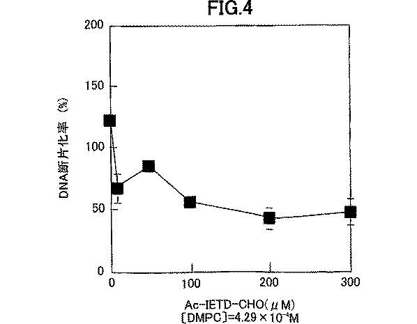
実施例1の1−1で調製したカスペース−9阻害剤のかわりに2−1で調製したプロカスペース−3分解酵素阻害剤を用いた以外は、実施例1の1−2と同様に行った。結果を図4に示した。
2−3.結果
図4から明らかなように、プロカスペース−3分解酵素阻害剤により、ほぼ濃度依存的なDNA断片化率の減少が観察されたことから、ジミリストイルホスファチジルコリン含有リポソーム液によるヒト前骨髄性白血病細胞のDNA断片化には、カスペース−3の活性化が関与することが示唆された。
実施例4
<フローサイトメーターによるカスペース−1,4阻害実験>
3−1.試薬の調製
・カスペース−1,4阻害剤(Ac−L−Tyr−L−Val−L−Ala−L−Asp−H、市販品、株式会社ペプチド研究所製、Ac−YVAD−CHOと略称することもある)は、5.7mgはかりとり、DMSO(ジメチルスルホキシド、市販品)382μlに溶解した。使用までサンプル容器の外部を乾燥させた状態で冷凍保存した(使用時は室温に戻した)。
3−2.カスペース阻害実験
実施例1の1−1で調製したカスペース−9阻害剤のかわりに3−1で調製したカスペース−1,4阻害剤を用いた以外は、実施例1の1−2と同様に行った。結果を図5に示した。
3−3.結果
図5から明らかなように、カスペース−1,4阻害剤により、ほぼ濃度依存的なDNA断片化率の減少が観察されたことから、ジミリストイルホスファチジルコリン含有リポソーム液によるヒト前骨髄性白血病細胞のDNA断片化には、カスペース−1または−4の活性化が関与することが示唆された。
実施例5
<ウェスタンブロット法によるカスペース−3基質分解物の検出>
4−1.ウェスタンブロッティング法によるカスペース−3基質分解物の検出について
DTTなどの還元剤を用いてジスルフィド結合を切断したタンパク質試料に、陰イオン界面活性剤であるSDSを添加することにより、タンパク質は変性され、タンパク質にSDSが定量的に結合することで、電気泳動によりタンパク質を分子量により分離することができる。分離したタンパク質をニトロセルロース膜に電気的にブロッティングし、カスペース−3の基質の一つであるpoly(ADP−ribose)polymerase(PARPと略称する)の分解物に特異的に結合する一次抗体(Anti−PARP p85 Fragment rabbit polyclonal antibodyを結合させた後、アルカリホスファターゼ標識した二次抗体(Donkey Anti−Rabbit IgG(H+L)Alkalie Phosphatase conjugate)を結合させ、これにアルカリホスファターゼの基質であるBCIP及びNBTを添加することで、BCIPのリン酸の切断とNBTの還元により不溶性の紫色の沈殿を生じ、抗体を検出することができる。
4−2.ウェスタンブロッティング法による基質分解物の検出試薬の一覧
・Sample Buffer(1.0M Tris−HCl(pH6.8),40%SDS,25%Glycerol,1%Bromophenol Blue,0.13M DTT)
・Trans Buffer(0.025M Tris,0.2M Glycine,10%Metanol)
・TBST(0.02M Tris,10%Tween20,0.05M NaCl)
・Blocking Buffer(TBST+5%スキムミルク)
・Anti−PARP p85 Fragment pAb(PROMEGA社製)
・Donkey Anti−Rabbit IgG(H+L)AP(PROMEGA社製)
・Tween20(PROMEGA社製)
・Glycine(amresco社製)
・メタノール特級(ナカライテスク株式会社製)
・スキムミルク(雪印社製)
・Sodium Laulyl Sulfate(ナカライテスク株式会社製)
・Blot Qualified BSA(PROMEGA社製)
・Tris(hydroxymethyl)aminomethane(ナカライテスク株式会社製、Trisと略称する)
・Bromophenol Blue(和光純薬株式会社製)
・Hydrochoric Acid(ナカライテスク株式会社製)
・Glycerol(GIBCO BRL社製)
・ジチオスレイトール(ナカライテスク株式会社製、DTTと略称する)
・Migration Buffer(BIO−RAD社製、10×Tris/Glycine/SDS buffer for SDS−PAGE applications)
・CBB R−250染色液(BIO−RAD社製)
・CBB R−250脱色液(BIO−RAD社製)
・Kaleidoscope prestained standards(PROMEGA社製)
・READY−GELS J for Polyacrylamide Electrophoresis(BIO−RAD社製)
・Western Blue Stabilized Substrate for Alkaline Phosphatase(PROMEGA社製)
4−3.ウェスタンブロッティング法によるカスペース−3基質分解物の検出実験操作
4−2の試薬を用いて次の実験を行った。まずヒト前骨髄性白血病細胞(HL−60、理化学研究所細胞銀行より購入したもの)の懸濁液(50×104cells/ml)12mlにジミリストイルホスファチジルコリン含有リポソーム液([DMPC]=1.5×10−2M,[C12(EO)10]=1.67×10−3M)を2ml添加し、3時間インキュベートした。これを2mlの−PBSで洗浄した後、2000ppmで6分間、遠心分離し、上澄みを除去した後、−PBS(400μl)及び5×Sample Buffer(100μl)を加えた。その後、ポリトロン(商品名POLYTRON PT3100、KINEMATICA AG社製)乳化器でヒト前骨髄性白血病細胞を破砕(15000rpm,5min)し、更に、バス型超音波照射機で37℃、10分間超音波照射してホモジネートを作製した。その後、100℃の湯浴で5分間暖め、15000rpmで30分間、遠心分離した。上澄みをwellに15μlづつ注入し、SDS−PAGE(200V,45min)によりタンパクを分離した後、ニトロセルロース膜にタンパク質をブロッティング(50V,50min)し、Blocking Bufferを用いて37℃で30分、ブロッキングを行った。これに一次抗体を転写面に添加し、室温で90分反応させた後、TBST bufferで洗浄し、続いて二次抗体を一次抗体結合面に添加し、室温で90分反応させた。その後、TBST bufferで洗浄し、更に、脱イオン水で洗浄した後、発色基質を添加し、PARP分解物の検出を行った。結果を図4に示した。
4−4.カスペース−3基質分解物の検出実験の結果
カスペース−3は、アポトーシスシグナルにおいてカスペースカスケード活性化の末端で活性化され、多くの基質を分解することによりアポトーシスを誘導する。4−3の実験により、カスペース−3の基質の一つであるPARPの分解物をウェスタンブロッティング法を用いて検出し、ジミリストイルホスファチジルコリン含有リポソーム液によるヒト前骨髄性白血病細胞内でのカスペース−3の活性化について検討できた。即ち、図6から明らかなように、ジミリストイルホスファチジルコリン含有リポソーム液で処理したものではPARP分解物が検出されたが、ジミリストイルホスファチジルコリン含有リポソーム液未添加においては検出されなかった。このことから、ジミリストイルホスファチジルコリン含有リポソーム液によりヒト前骨髄性白血病細胞内で、カスペース−3が活性化されることが示唆された。
実施例6
<DMPCのメラノーマに対する抗腫瘍実験>
5−1.DMPC分散液の調製
DMPC(商品名コートソームMC−4040、市販品、日本油脂株式会社製)を所定量秤量し、緩衝水溶液として−PBSを加え、バス型超音波照射器(商品名BRANSONIC MODEL B2210:90W)で45℃、1ml/1minの条件で超音波照射し、更に得られた溶液を0.45μmフィルター(商品名ADVANTEC DISMC−13cp)で濾過してDMPC分散液を調製した。尚、このときDMPC秤量時の量をかえることによりDMPCの濃度が、5.0×10⁻²M、5.0×10⁻³M、5.0×10⁻⁴Mの3種のDMPC分散液を調製した。
5−2.担癌マウスを用いたin vivo治療実験
動物は、C57BL/6雌性マウス(5週齢、日本チャールスリバーより購入)を5日間の検疫馴化後、6週齢より1群6匹で実験に使用した。飼育環境は、室温24±2℃、湿度55±10%で、水及び餌は自由に摂取させた。
マウスは癌細胞移植当日の体重をもとに層別連続無作為化法により1群6匹づつに群わけを行った。B16−F0メラノーマ細胞(5.0×10⁵cells)をマウスの腹腔内に移植し、担癌マウスを作成した。DMPC分散液(5−1で調製したもの)の投与方法は、腹腔内投与とし、実験スケジュールは、癌移植当日(移植1時間後)から12日目まで計13回投与(20ml/kg体重)を行う実験を行った。投与終了後は、全ての動物が死亡するまで観察を行い、マウスの生存日数を測定し、治療群の平均生存日数を未治療群の平均生存日数で除して得た値に100を剰じて、延命率(%)を算出した。結果を図7に示し、治療効果を検討した。
以上の結果から、DMPC分散液は、担癌マウスに対して顕著な治療効果を持つことが明かとなった。DMPC単一成分の分散液が、このような癌治療効果を示すことはこれまで報告がなく、驚くべきことである。従って、新しい発見として、リン脂質の一種であるDMPCが、高い癌治療効果を有することが初めて明かとなり、まったく副作用のない新しい癌治療薬として期待できることがわかった。
本発明アポトーシス誘導剤は、通常の状態ではアポトーシスのメカニズムが正常に機能しない腫瘍細胞に、アポトーシスを誘導することができる。また、本発明のカスペースカスケード活性化剤は、通常の状態ではアポトーシスのメカニズムが正常に機能せず、従ってカスペースカスケードも活性化していない腫瘍細胞に、アポトーシスを誘導することによりカスペースカスケードを活性化させ、アポトーシスを実行することができる。
更に、本発明の制癌剤は、副作用のない安全な制癌剤として極めて有効である。
【図面の簡単な説明】
図1は、ハイブリッドリポソームにより処理したHL−60細胞由来のDNAのアガロースゲル電気泳動である。
図中、Aはコントロールであり;BはDMPCであり;CはC12(EO)4であり;DはDMPC/10モル%C12(EO)4であり;EはC12(EO)10であり;FはDMPC/10モル%C12(EO)10であり;GはC12(EO)12であり;HはDMPC/10モル%C12(EO)12であり;IはC12(EO)23であり;JはDMPC/10モル%C12(EO)23であり;KはDNAマーカーである。
図2は、DMPC/10モル%C12(EO)10のハイブリッドリポソームで処理したHL−60細胞の蛍光顕微鏡観察である。
図3は、ジミリストイルホスファチジルコリン含有リポソーム液によるカスペース−9阻害効果を示す図である。
図4は、ジミリストイルホスファチジルコリン含有リポソーム液によるカスペース−3阻害効果を示す図である。
図5は、ジミリストイルホスファチジルコリン含有リポソーム液によるカスペース−1/−4阻害効果を示す図である。
図6は、ウエスタンブロット法によるカスペース−3基質分解物の検出を示す図である。DMPCとC12(EO)10とのハイブリッドリポソームで処理したHL−60細胞のウェスタン分析である。
図7は、DMPC分散液による担癌マウスの治療実験結果を示す図である。B16−F0メラノーマ細胞を接種後、DMPCリポソームで処理(0〜12日)したマウスの生存曲線である。
図中、●はコントロール、△は[DPMC]=5.0×10⁻²M、■は[DPMC]=5.0×10⁻³M、◇は[DPMC]=5.0×10⁻⁴Mを表す。Background of the Invention
The present invention relates to a novel apoptosis inducer, a caspase cascade activator, and an anticancer agent.
The concept of apoptosis was first proposed by Kerr, Wylli. Currie, 1972 (Kerr JFR, Wyllie AH and Currie AR, Br. J. Cancer, 26, 239-257 (1972)). Apoptosis is characterized by significant morphological changes, such as cell shrinkage, chromatin condensation, and fragmentation of DNA into nucleosome units (approximately 180 bp), and necrosis (necrosis) is sudden cell death due to the physical action of an external stimulus. Whereas, defined as genetically controlled cell death for individual maintenance.
The mechanism of apoptosis is thought to go through three pathways: induction, decision and execution. The decision process that plays a major role in the mechanism of apoptosis has two signaling pathways. One is the mitochondrial-mediated pathway, which is regulated by the Bcl-2 family. The other is a so-called Kaspace Cascade, which is a self-decomposition by Kaspace and a transmission path by limited decomposition of Kaspace under the Kaspace cascade.
The molecular mechanism of this apoptosis was examined by Horvitz, Yuan et al. Using nematodes. They discovered a total of 14 genes involved in apoptosis of cells constituting nematodes, and found that ced-3 and ced-4 were particularly important as apoptosis-executing factors and ced-9 was a suppressor. As a result of gene identification, it was found that ced-3 had high homology to a cysteine protease called human IL-1β converting enzyme (ICE) (Yuan J., Shaham S., Ledoux S., Ellis H.M. and Horvitz). HR, Cell, 75, 641-652 (1993)). Furthermore, since it was confirmed that apoptosis was induced by overexpression of ICE, it became possible that ICE might also act as an apoptosis-executing factor in humans. However, the subsequently reported ICE knockout mice had little effect on apoptotic function. It was also later shown that the degradation of poly (ADP-ribose) polymerase, a characteristic of apoptosis, does not occur with ICE, but with a cysteine protease similar to ICE. From these, it was speculated that the apoptosis-executing factor in humans was not ICE itself but another ICE-like enzyme, and it was considered that ICE-like enzymes other than ICE exist in humans.
From the above, identification of ICE-like enzymes was attempted since the report of Yuan et al., And it was found that there are about 10 types of ICE-like enzymes in humans including ICE. The identified ICE-like enzyme is an Asp-specific cysteine protease, and the enzyme name is unified to “casspace”, and the numbers are given in the order of reporting, such as caspase-1 (ICE) to caspase-10. As a result, it has come to be generally referred to as the caspase family protease.
At present, caspases are roughly classified into three groups based on their substrate specificity, and group I centered on caspase-1 is thought to be involved in the production and secretion of interleukins, and is centered on caspase-3. Group II is considered to be the center of apoptosis-executing molecules, and group III represented by caspase-8 / -9 is located upstream of the caspase proteolytic cascade, and functions to transmit cell death signals for apoptosis. It is believed that there is.
The caspase family has an activity of cleaving the C-terminal side of Asp (D) by a common active center sequence, and has slightly different substrate specificities. In general, caspase is formed as an inactive precursor and is cleaved into two subunits by an activating enzyme to form an active caspase. The cleavage site in activation of the precursor is determined by Asp in the caspase molecule. It is on the C-terminal side of the residue and coincides with the substrate recognition site of caspase. Therefore, it is presumed that the mechanism for activating the kaspace itself is working. In the apoptosis induced by Fas, the phenomenon that caspase-3 like activity is increased after activation of caspase-1 is observed, and that caspase-3 is located downstream of caspase-1. Is known (Enari M., Talanian RV, Wong WWW and Nagata S., Nature, 380, 723-726 (1996)). This suggests that during the execution of apoptosis, several caspases are continuously activated, forming a so-called caspase cascade. Caspase also plays an important role in the process of executing apoptosis by degrading intracellular proteins.
As described above, it is interesting that a large number of kaspaces exist in humans, and it is considered that a complex kaspace network is formed in order to maintain individuals.
However, the mechanism by which the kaspace cascade is actually activated remains largely unknown at present.
As described above, apoptosis and the caspase cascade, which play important roles in vivo, are also important in drug development, and by utilizing the apoptosis and caspase cascade mechanism, the apoptosis induction mechanism can function normally. It is contemplated to develop therapeutics for cell proliferative disorders, such as cancers that do not.
For example, JP-A-2000-143532 discloses that a cell proliferative disease is treated by activating a caspase cascade and inducing apoptosis with a drug having a cyclic RGD sequence as a pharmacologically active ingredient. . However, an apoptosis inducer and a caspase cascade activator containing dimyristoyl phosphatidylcholine as an active ingredient have not been known so far.
On the other hand, some anticancer agents using a lipid component are known. In general, most anticancer drugs using lipid components are in the form of liposomes or emulsions and use lipid components as carriers for transporting drugs, and few lipid components themselves act as anticancer agents. Publications showing the effectiveness of lipid components against cancer are listed below. JP-A-6-256181 discloses a cell differentiation inducer containing tocopherol as an active ingredient, and describes that it is effective as a therapeutic or ameliorating agent for cancer. JP-A-1-203330 describes an anticancer drug containing docosahexaenoyl-lysophosphatidylcholine as an active ingredient. JP-A-1-203322 describes an anticancer drug containing docosahexaenoyl monoglyceride as an active ingredient. JP-A-1-203331 describes a carcinostatic agent comprising 1,2-didocosahexaenoyl-phosphatidylcholine as an active ingredient. Japanese Unexamined Patent Publication (Kokai) No. Hei 3-163031 describes a liquid membrane anticancer agent obtained by adding an anticancer agent to a mixed molecular assembly formed by a phospholipid and a micelle surfactant. JP-A-2-11516 discloses an anticancer agent containing one type of phospholipid as an active ingredient. With respect to known anticancer drugs utilizing phospholipids, the mechanism of their anticancer activity has not been elucidated at all, and even less studies have been known that antitumor activity is attributed to apoptosis.
Disclosure of the invention
A first object of the present invention is to provide a novel apoptosis inducer containing di (alkylcarbonyl) phosphatidylcholine or the like as an active ingredient.
A second object of the present invention is to provide a novel caspase cascade activator containing di (alkylcarbonyl) phosphatidylcholine or the like as an active ingredient.
A third object of the present invention is to provide an anticancer drug which utilizes di (alkylcarbonyl) phosphatidylcholine or the like as an active ingredient and has no side effects.
The present inventors have noted glycerophospholipids such as dimyristoyl phosphatidylcholine well known as a kind of phospholipids, and surprisingly found that these compounds act as an apoptosis inducer, and an activator of caspase cascade, The present invention has been completed based on the new finding that it can be used as a highly safe anticancer drug without side effects as compared with conventional anticancer drugs.
That is, the present invention provides, as an active ingredient, at least one selected from the group consisting of di (alkylcarbonyl) phosphatidylcholine, di (alkylcarbonyl) phosphatidylalkanolamine, di (alkylcarbonyl) phosphatidylinositol, and di (alkylcarbonyl) phosphatidylserine. An apoptosis-inducing agent characterized by comprising:
The present invention also contains, as an active ingredient, at least one selected from the group consisting of di (alkylcarbonyl) phosphatidylcholine, di (alkylcarbonyl) phosphatidylalkanolamine, di (alkylcarbonyl) phosphatidylinositol, and di (alkylcarbonyl) phosphatidylserine. To provide a caspase cascade activator.
The present invention also provides a carcinostatic agent characterized by causing tumor cells to undergo apoptosis by using these apoptosis inducers or caspase cascade activators.
The present invention also provides an anticancer agent characterized by apoptosis of tumor cells by activating the caspase cascade of tumor cells with a molecular assembly containing di (alkylcarbonyl) phosphatidylcholine or the like as an active ingredient.
The present invention further provides an anticancer drug comprising dimyristoyl phosphatidylcholine and a pharmaceutically acceptable carrier and / or adjuvant.
BEST MODE FOR CARRYING OUT THE INVENTION
As used herein, "apoptotic" refers to morphology in medicine and biology characterized by significant morphological changes such as cell shrinkage, chromatin condensation, DNA fragmentation at nucleosome units (about 180 bp). It is the process of causing a cell to undergo a defined, biochemically defined change, which leads to a programmed cell death different from necrosis (necrosis). Whether the cells are apoptotic can be confirmed by a known method such as observation of a DNA ladder by electrophoresis or morphological observation by an optical microscope.
As used herein, “activating the caspase cascade” refers to an apoptotic signal constructed in a cell by an Asp-specific cysteine protease (an enzyme that hydrolyzes the C-terminal of aspartic acid) called caspase. Activating the caspase cascade by bringing one of the caspases to an activated state in the transmission network, which necessarily activates all kinds of caspases in the cell. It does not mean. Which of the various caspases is activated may be different depending on the type and condition of the cell.
Activation of caspase can be estimated and confirmed by using a known method such as an inhibition experiment with a flow cytometer using a caspase inhibitor or detection of caspase degradation product by Western blotting.
Further, in the present specification, “tumor cells” are cells constituting so-called cancer or the like, or cells derived or related thereto, and are not particularly limited as long as they are cells generally called tumor cells. In addition, the disease is limited to human cells, including not only cells in organ tissues as a disease, but also cultured cells commonly used as test cells, and hybridomas derived from these tumor cells, etc. Not necessarily. Examples of such tumor cells include, for example, acute leukemia, chronic leukemia, malignant lymphoma, multiple myeloma, hematopoietic tumors such as macroglobulinemia, brain tumor, head and neck cancer, breast cancer, lung cancer, esophageal cancer, gastric cancer, Colorectal cancer, liver cancer, gallbladder / bile duct cancer, pancreatic cancer, pancreatic islet cell cancer, renal cell carcinoma, adrenocortical cancer, bladder cancer, prostate cancer, testicular tumor, ovarian cancer, uterine cancer, choriocarcinoma, thyroid cancer, malignant carcinoid tumor, skin Cells that form solid tumors such as cancer, malignant melanoma, osteosarcoma, soft tissue sarcoma, neuroblastoma, Wilms tumor, fetal rhabdomyosarcoma, and retinoblastoma.
As used herein, the term "pharmaceutically acceptable carrier and / or adjuvant" refers to a non-toxic carrier or a non-toxic carrier that can be administered to a patient together with dimyristoyl phosphatidylcholine and does not impair the pharmacological activity of dimyristoyl phosphatidylcholine. Represents an adjuvant.
First, the first embodiment of the present invention will be described.
In the first embodiment of the present invention, the active ingredient is selected from the group consisting of di (alkylcarbonyl) phosphatidylcholine, di (alkylcarbonyl) phosphatidylalkanolamine, di (alkylcarbonyl) phosphatidylinositol, and di (alkylcarbonyl) phosphatidylserine. The present invention relates to an apoptosis-inducing agent containing at least one.
The apoptosis-inducing agent of the present invention is effective for preventing or treating various diseases caused by the suppression of normal apoptotic function. Such diseases include cell proliferative diseases, malformations, autoimmune diseases and the like. In particular, it is effective for preventing or treating cell proliferative diseases such as cancer by apoptosis of tumor cells constituting cancer or the like in which the mechanism of apoptosis is directly or indirectly involved.
Therefore, the present invention also provides a compound selected from the group consisting of di (alkylcarbonyl) phosphatidylcholine, di (alkylcarbonyl) phosphatidylalkanolamine, di (alkylcarbonyl) phosphatidylinositol, and di (alkylcarbonyl) phosphatidylserine as an active ingredient. , A prophylactic or therapeutic agent for any of cell proliferative diseases, malformations or autoimmune diseases.
The present invention is also selected from the group consisting of di (alkylcarbonyl) phosphatidylcholine, di (alkylcarbonyl) phosphatidylalkanolamine, di (alkylcarbonyl) phosphatidylinositol, and di (alkylcarbonyl) phosphatidylserine for producing an apoptosis inducer. Related to the use of the compounds.
The present invention also provides a compound selected from the group consisting of di (alkylcarbonyl) phosphatidylcholine, di (alkylcarbonyl) phosphatidylalkanolamine, di (alkylcarbonyl) phosphatidylinositol, and di (alkylcarbonyl) phosphatidylserine as an active ingredient. To induce apoptosis in tumor cells by contacting the cells.
The present invention also provides a cell selected from the group consisting of di (alkylcarbonyl) phosphatidylcholine, di (alkylcarbonyl) phosphatidylalkanolamine, di (alkylcarbonyl) phosphatidylinositol, and di (alkylcarbonyl) phosphatidylserine as an active ingredient. The present invention relates to a method for preventing or treating these diseases, which comprises administering to a patient suffering from a proliferative disease, malformation or an autoimmune disease.
The two alkylcarbonyls contained in the active ingredient used in the present invention may be the same or different, but are preferably the same. The carbon number of the alkylcarbonyl group is preferably from 10 to 20, more preferably from 10 to 18, still more preferably from 12 to 16, and particularly preferably 14. Further, the alkyl group portion in the alkylcarbonyl group may be linear or branched.
When the active ingredient used in the present invention is di (alkylcarbonyl) phosphatidylalkanolamine, the alkanol moiety preferably has 1 to 6 carbon atoms, more preferably 1 to 3 carbon atoms, and particularly preferably 2 carbon atoms.
A preferred active ingredient used in the present invention is represented by the following general formula (1).
R 2 Is OCH 2 CH 2 N + (CH 3 ) 3 , OCH 2 CH 2 NH 2 , OCH 2 CH (NH 2 ) COOH or the following formula (2)
Is a group represented by
As the active ingredient used in the present invention, di (alkylcarbonyl) phosphatidylcholine is preferable.
The most preferred compound represented by the general formula (1) is dimyristoylphosphatidylcholine (DMPC) represented by the following general formula (3).
Note that dimyristoyl phosphatidylcholine is one of phospholipids called phosphatidylcholine or lecithin, and is formally referred to as 1,2-dimyristoyl-sn-glycero-3-phosphocholine. . Dimyristoyl phosphatidylcholine is sometimes referred to as dimyristoyl lecithin or L-α-dimyristoyl phosphatidylcholine.
The active ingredients used in the present invention can be used alone or in combination of two or more. When two or more kinds are used in combination, it is preferable to use di (alkylcarbonyl) phosphatidylcholine, particularly DMPC, in combination with other active ingredients.
The active ingredient such as di (alkylcarbonyl) phosphatidylcholine used in the present invention may be derived from a natural product or a chemically synthesized product. When it is derived from a natural product, it can be obtained by extracting, separating, and purifying from raw materials such as animals, plants, and microorganisms, such as brain, liver, egg yolk, soybean, and yeast, by known methods. These active ingredients can also be obtained by using a commercially available lecithin as a raw material and utilizing a known separation method utilizing a difference in fatty acid chain length. When derived from a chemically synthesized product, these active ingredients can be easily synthesized by making full use of known synthetic chemistry knowledge.For example, in the case of dimyristoyl phosphatidylcholine, myristic acid is used as a glycerophosphocholine. It can also be obtained by a method such as introduction by a method. In addition, commercially available products may be used, and those having high purity of a pharmaceutical grade are particularly preferable. For example, DMPC is commercially available from NOF Corporation under the trade name of Coatsome MC-4040.
The apoptosis-inducing agent of the present invention is obtained by mixing an active ingredient with a pharmacologically acceptable carrier known per se, an excipient, a disintegrating agent, a corrective agent, a bulking agent, a diluent, a solubilizing agent, and the like. In the form of pharmaceutical compositions, for example, tablets, capsules, granules, powders, powders, pills, solvents, drinks, injections, drops, suppositories and the like. Such formulations can be administered orally or parenterally.
The dose when the apoptosis-inducing agent of the present invention is used for the treatment of disease and the like varies depending on the administration subject, administration route, symptoms and the like. -1 ~ 1 × 10 -6 It is preferable to administer 0.01 to 100 ml / kg body weight of a solution containing M concentration about 1 to 3 times a day. 5 × 10 active ingredients -2 ~ 5 × 10 -4 More preferably, 1 to 50 ml / kg body weight of a solution containing M concentration is administered about 1 to 3 times a day.
Next, a second embodiment of the present invention will be described.
In a second aspect of the present invention, at least one selected from the group consisting of di (alkylcarbonyl) phosphatidylcholine, di (alkylcarbonyl) phosphatidylalkanolamine, di (alkylcarbonyl) phosphatidylinositol, and di (alkylcarbonyl) phosphatidylserine is used as an active ingredient. A caspase cascade activator containing one.
The activator of caspase cascade of the present invention is effective for prevention or treatment of various diseases caused by suppression of the function of normal caspase cascade activation. Such diseases include cell proliferative diseases, malformations, autoimmune diseases and the like. In particular, activating the caspase cascade promotes the normalization of the apoptotic mechanism, which is effective for preventing or treating cell proliferative diseases such as tumor cells constituting cancer and the like.
Therefore, the present invention also provides di (alkylcarbonyl) phosphatidylcholine, di (alkylcarbonyl) phosphatidylalkanolamine, di (alkylcarbonyl) phosphatidylinositol, di (alkylcarbonyl) phosphatidylserine for producing a caspase cascade activator. The use of a compound selected from the group consisting of:
The present invention also provides a compound selected from the group consisting of di (alkylcarbonyl) phosphatidylcholine, di (alkylcarbonyl) phosphatidylalkanolamine, di (alkylcarbonyl) phosphatidylinositol, and di (alkylcarbonyl) phosphatidylserine as an active ingredient. For activating the caspase cascade of tumor cells by contacting the cell with the cell.
The active ingredient used as the cascade activator of the present invention is the same as that described for the apoptosis inducer.
The caspase cascade activator of the present invention is obtained by mixing the active ingredient with a pharmacologically acceptable carrier, excipient, disintegrating agent, corrective agent, bulking agent, diluent, solubilizing agent and the like known per se. According to a known method, it can be formulated into a pharmaceutical composition such as a tablet, capsule, granule, powder, powder, pill, solvent, drink, injection, infusion, suppository and the like. Such formulations can be administered orally or parenterally.
When the caspase cascade activator of the present invention is used for treating a disease or the like, the dose varies depending on the administration subject, administration route, symptoms, and the like. 1 × 10 active ingredients -1 ~ 1 × 10 -6 It is preferable to administer 0.01 to 100 ml / kg body weight of a solution containing M at a concentration of about 1 to 3 times a day. 5 × 10 active ingredients -2 ~ 5 × 10 -4 More preferably, 1 to 50 ml / kg body weight of a solution containing M concentration is administered about 1 to 3 times a day.
Finally, a third embodiment of the present invention will be described.
According to a third aspect of the present invention, at least one selected from the group consisting of di (alkylcarbonyl) phosphatidylcholine, di (alkylcarbonyl) phosphatidylalkanolamine, di (alkylcarbonyl) phosphatidylinositol, and di (alkylcarbonyl) phosphatidylserine is used as an active ingredient. The present invention relates to a type of anticancer drug. The active ingredient used as the anticancer agent of the present invention is the same as that described for the apoptosis inducer.
The anticancer agent of the present invention, by effectively acting on tumor cells and the like in which the function of activating the normal caspase cascade is suppressed, activates the caspase cascade and apoptosis of the tumor cells and the like. Cancer can be prevented or treated. The target disease of the anticancer agent of the present invention is not limited to these as long as it is a so-called cancer, but the above-mentioned cancers classified as hematopoietic tumors and solid tumors, particularly acute leukemia, chronic leukemia, malignant melanoma, lung cancer, liver cancer The anticancer agent of the present invention is particularly excellent for preventing or treating gastric cancer and malignant brain tumor.
Therefore, the present invention also provides a compound selected from the group consisting of di (alkylcarbonyl) phosphatidylcholine, di (alkylcarbonyl) phosphatidylalkanolamine, di (alkylcarbonyl) phosphatidylinositol, and di (alkylcarbonyl) phosphatidylserine as an active ingredient. And a preventive or therapeutic agent for acute leukemia, chronic leukemia, malignant melanoma, lung cancer, liver cancer, stomach cancer, and malignant brain tumor.
The present invention also relates to a compound selected from the group consisting of di (alkylcarbonyl) phosphatidylcholine, di (alkylcarbonyl) phosphatidylalkanolamine, di (alkylcarbonyl) phosphatidylinositol and di (alkylcarbonyl) phosphatidylserine for producing an anticancer agent. About use.
The present invention also provides a compound selected from the group consisting of di (alkylcarbonyl) phosphatidylcholine, di (alkylcarbonyl) phosphatidylalkanolamine, di (alkylcarbonyl) phosphatidylinositol, and di (alkylcarbonyl) phosphatidylserine as an active ingredient. The present invention relates to a method for preventing or treating leukemia, chronic leukemia, malignant melanoma, lung cancer, liver cancer, stomach cancer, or malignant brain tumor, including administering to a patient having any of these diseases.
The anticancer agent of the present invention comprises at least one selected from the group consisting of di (alkylcarbonyl) phosphatidylcholine, di (alkylcarbonyl) phosphatidylalkanolamine, di (alkylcarbonyl) phosphatidylinositol, and di (alkylcarbonyl) phosphatidylserine, which is known per se. Pharmacologically acceptable carriers, excipients, disintegrants, correctors, extenders, diluents, diluents, mixed with solubilizers and the like, and according to known methods, pharmaceutical compositions such as tablets, capsules, granules, It can be freely formulated into powders, powders, pills, solvents, suppositories and the like. Such formulations can be administered orally or parenterally. In particular, it is preferably used as a drink, injection or infusion.
When administered as an injection, infusion, or drink, it is preferable to administer an active ingredient such as di (alkylcarbonyl) phosphatidylcholine in a molecular assembly. Examples of molecular assemblies containing these active ingredients include fat emulsions, polymer micelles, complexes with proteins such as albumin, liposomes, and the like. Liposomes are most preferred because they have a forming function.
When forming liposomes, the active ingredient concentration is 5 × 10 -4 ~ 1.0 × 10 -1 M, preferably 1 × 10 -2 ~ 5 × 10 -2 M is more preferable.
The liposome in the present invention may be formed by any known method. For example, known production methods such as a thin film method, a reverse phase evaporation method, a freeze-thaw method, an ethanol injection method, a high-pressure emulsification method, an ultrasonic dispersion method, a dialysis method, and an extrusion method can be appropriately used. The method described in JP-A-9-87168 may be used.
As the aqueous solvent that can be used for preparing the liposome, simple water may be used, or an aqueous mixed solvent containing a small amount of ethanol or the like, or plasma may be used. Or distilled water or ultrapure water. Known physiologically active substances, proteins, buffer substances, various salts, plasma, sugars, and the like may be dissolved in an aqueous solvent to prepare a solution.
The liposome obtained in this manner may be purified by a method such as ultrafiltration, centrifugation, gel filtration, or the like, and operations such as concentration and dilution may be freely performed.
The liposome of the present invention may contain known liposome components to the extent that the purpose of the present invention is not violated. Specific examples include phospholipids such as egg yolk lecithin, soybean lecithin, hydrogenated egg yolk lecithin, hydrogenated soybean lecithin, and phosphatidylglycerol.
Although a small amount of cholesterol can be used as a membrane-stabilizing component of the liposome, a large amount of cholesterol is not preferred because it may reduce the antitumor activity of dimyristoylphosphatidylcholine. Further, phosphatidic acid or fatty acid may be added as a charged substance.
Further, as a dispersion stabilizer, a surfactant known by trade names such as alkyl PEG having 10 to 20 carbon atoms, represented by polyoxyethylene dodecyl ether, and Triton and Pluronic can be added. Among them, alkyl PEG having 12 to 14 carbon atoms is preferable. In particular, it is preferable to use a hybrid liposome formed from di (alkylcarbonyl) phosphatidylcholine and a surfactant that forms micelles. Among them, it is preferable to combine DMPC with an alkyl PEG having 10 to 16 carbon atoms and 2 to 30 EO (preferably having 12 to 14 carbon atoms and 2 to 24 EO).
In the present invention, a saccharide can be particularly preferably added to an aqueous solvent for the purpose of contributing to the stabilization of the dispersion of the liposome and enhancing the anticancer activity. Known saccharides such as monosaccharides, disaccharides, oligosaccharides, and polysaccharides can be used freely without particular limitation depending on the purpose, and sucrose and trehalose are particularly preferable. Also, the amount of addition may be appropriately selected. Further, α-tocopherol or the like may be added to the liposome as an antioxidant.
The diameter of the liposome to be prepared may be any diameter as long as it is within a medically acceptable range, but is preferably about 20 nm to 1 μm, more preferably about 30 nm to 300 nm, and still more preferably about 30 nm to 300 nm. For example, the thickness is preferably about 40 nm to 180 nm. The liposome is preferably prepared so as to be uniformly dispersed in an aqueous solvent, and when precipitation or aggregation is observed, it is not preferable because it is difficult to use the liposome as a carcinostatic agent mainly for physical reasons.
When the anticancer agent of the present invention is used for treating a disease or the like, the dose varies depending on the administration subject, administration route, symptoms, and the like. -1 ~ 1 × 10 -6 M solution having a concentration of 0.01 to 100 ml / kg body weight, preferably 5 × 10 -2 ~ 5 × 10 -4 It is preferable to administer 1 to 50 ml / kg body weight of a solution containing M at a concentration of about 1 to 3 times a day.
Example
Hereinafter, the present invention will be described more specifically with reference to Examples, but the present invention is not limited thereto.
Example 1
<Apoptosis induction experiment using liposomes>
1-1. Preparation of liposomes
The hybrid liposome solution was prepared by adding a predetermined amount of dimyristoyl phosphatidylcholine (trade name: coatsome MC-4040, manufactured by NOF Corporation) and polyoxyethylene dodecyl ether (commercially available, manufactured by Sigma) in an eggplant-shaped flask containing PBS. The sample was weighed and irradiated with ultrasonic waves by a bath-type ultrasonic irradiator (trade name: BRANSONIC MODEL B2210: 90W) under the conditions of 45 ° C. and 1 ml / 1 min, and the obtained solution was further filtered with a 0.45 μm filter (trade name, ADVANTEC DISMC). -13 cp).
1-2. Observation result of DNA ladder by electrophoresis and observation of cell fragmentation by fluorescence microscope
Using the hybrid liposome prepared in 1-1, human promyelocytic leukemia cells (HL-60, purchased from RIKEN Cell Bank) were processed, and the DNA ladder was observed by agarose gel electrophoresis of the DNA. did. In addition, cell fragmentation was observed with a fluorescence microscope using the fragmented DNA-staining method. The results are shown in FIGS. 1 and 2, respectively.
Example 2
<Caspase-9 inhibition experiment using flow cytometer>
1-1. Preparation of reagents
-The RNase solution was prepared by dissolving RNase (commercially available, manufactured by amresco) in -PBS so as to have a concentration of 0.25 mg / ml, and stored frozen until use (returned to room temperature when used). .
-The PI solution was prepared by dissolving Propidium Iodide (commercial product, manufactured by Funakoshi Co., Ltd., abbreviated as PI) in -PBS to a concentration of 0.5 mg / ml. It was stored in a refrigerator protected from light until use (when used, it was returned to room temperature).
-70% ethanol was obtained by mixing 99% ethanol (commercial product, manufactured by Nacalai Tesque, Inc.) and -PBS at a ratio of 7: 3. Stored frozen until use (returned to room temperature during use).
-Caspase-9 inhibitor (Ac-L-Leu-L-Glu-L-His-L-Asp-H, a commercial product, manufactured by Peptide Research Institute, Inc., and may be abbreviated as Ac-LEHD-CHO) Was weighed in an amount of 11.0 mg and dissolved in 404 μl of DMSO (dimethyl sulfoxide, commercially available). The sample container was frozen and stored in a dry state until use (the temperature was returned to room temperature during use).
The hybrid liposome solution was dimyristoyl phosphatidylcholine (trade name: Coatsome MC-4040, commercially available product, manufactured by NOF Corporation, sometimes abbreviated as DMPC) at 1.5 × 10 5 -2 M and the dispersant polyoxyethylene dodecyl ether (C 12 (EO) 10 (Commercial product, manufactured by Sigma) 1.67 × 10 -3 M was weighed into an eggplant-shaped flask containing a buffered aqueous solution -PBS so as to have a concentration of M, and was superposed at 45 ° C. for 1 ml / 1 min with a bath-type ultrasonic irradiator (trade name: BRANSONIC MODEL B2210: 90 W). The solution was irradiated with a sound wave, and the obtained solution was filtered through a 0.45 μm filter (trade name: ADVANTEC DISMC-13cp) to prepare a solution. The particle size of the thus obtained dimyristoyl phosphatidylcholine-containing liposome solution was measured by a dynamic light scattering method using an electrophoretic light scattering photometer (trade name, Photo ELS-8000, manufactured by OTUKA ERECTRONICS). The diameter of the liposome was about 76 nm, and it was confirmed that the liposome was stable for more than one month in the measurement with time.
1-2. Caspase inhibition experiment
The following experiment was performed using the reagent prepared in 1-1. First, a suspension of human promyelocytic leukemia cells (HL-60, purchased from RIKEN Cell Bank) (50 × 10 4 (cells / ml) A plurality of samples were prepared by adding 0 to 120 μl of caspase-9 inhibitor stepwise to 12 ml, and after incubating for 90 minutes, 2 ml of liposome solution containing dimyristoyl phosphatidylcholine was added to each sample, and further incubated for 3 hours did. After washing with 2 ml of -PBS, 5 ml of 70% ethanol was added, and the mixture was allowed to stand at ice temperature for 1 hour to perform tissue fixation. After washing with 2 ml of -PBS, 6 ml of RNase solution was added and incubated for 30 minutes. Next, 670 μl of the PI solution was added, light-shielded with aluminum foil, and left for 30 minutes in a dark place at an ice temperature to perform PI staining. After PI staining, the mixture was centrifuged at 2000 rpm for 5 minutes and the supernatant was removed. The precipitate was suspended in 500 µl of -PBS, and the DNA content was measured by a flow cytometer (trade name: COULTER EPICS-XL).
The inhibitory effect was determined by comparing the DNA fragmentation rate of the sample to which the caspase-9 inhibitor was added stepwise with respect to the DNA fragmentation rate of the control (to which only the liposome solution containing dimyristoylphosphatidylcholine was added without adding the caspase-9 inhibitor). The results were shown in FIG.
1-3. result
As is apparent from FIG. 3, an almost concentration-dependent decrease in the DNA fragmentation rate was observed with the caspase-9 inhibitor. Therefore, the DNA fragment of human promyelocytic leukemia cells with the liposome solution containing dimyristoylphosphatidylcholine was observed. It was suggested that the activation involved the activation of caspase-9.
In the control to which only the liposome solution containing dimyristoyl phosphatidylcholine was added without adding the caspase-9 inhibitor, clear DNA fragmentation was observed in human promyelocytic leukemia cells, and it was confirmed that apoptosis was induced. Was.
Example 3
<Procaspace-3 degrading enzyme inhibition experiment using a flow cytometer>
2-1. Preparation of reagents
-Procaspace-3 degrading enzyme inhibitor (Ac-L-Ile-L-Glu-L-Thr-L-Asp-H, a commercial product, abbreviated as Ac-IETD-CHO, manufactured by Peptide Research Institute Co., Ltd.) 5.4 mg was weighed and dissolved in 355 μl of DMSO (dimethyl sulfoxide, commercially available product). The sample container was frozen and stored in a dry state until use (the temperature was returned to room temperature during use).
2-2. Caspase inhibition experiment
In the same manner as in 1-2 of Example 1, except that the procaspace-3 degrading enzyme inhibitor prepared in 2-1 was used instead of the caspase-9 inhibitor prepared in 1-1 in Example 1, went. The results are shown in FIG.
2-3. result
As is evident from FIG. 4, since the concentration of DNA fragmentation was substantially reduced in a concentration-dependent manner by the procaspace-3 degrading enzyme inhibitor, human promyelocytic leukemia cells were treated with dimyristoyl phosphatidylcholine-containing liposome solution. It was suggested that the activation of caspase-3 was involved in the DNA fragmentation of Caspase-3.
Example 4
<Caspase-1,4 inhibition experiment using flow cytometer>
3-1. Preparation of reagents
Caspase-1,4 inhibitor (Ac-L-Tyr-L-Val-L-Ala-L-Asp-H, a commercial product, manufactured by Peptide Research Institute, Inc., and may be abbreviated as Ac-YVAD-CHO. 5.7 mg was weighed and dissolved in 382 μl of DMSO (dimethyl sulfoxide, commercially available). The sample container was frozen and stored in a dry state until use (the temperature was returned to room temperature during use).
3-2. Caspase inhibition experiment
Performed in the same manner as 1-2 in Example 1, except that the caspase-1,4 inhibitor prepared in 3-1 was used instead of the caspase-9 inhibitor prepared in 1-1 of Example 1 Was. The results are shown in FIG.
3-3. result
As is evident from FIG. 5, a substantially concentration-dependent decrease in the rate of DNA fragmentation was observed with the caspase-1,4 inhibitor. Thus, the liposome solution containing dimyristoyl phosphatidylcholine was used to induce the conversion of human promyelocytic leukemia cells. It was suggested that DNA fragmentation involves activation of caspase-1 or -4.
Example 5
<Detection of Kaspace-3 Substrate Degradation Products by Western Blot Method>
4-1. Detection of caspase-3 substrate degradation products by Western blotting
By adding SDS, which is an anionic surfactant, to a protein sample in which disulfide bonds have been cleaved using a reducing agent such as DTT, the protein is denatured, and SDS is quantitatively bound to the protein. Can separate proteins by molecular weight. The separated protein is electrically blotted on a nitrocellulose membrane, and a primary antibody (specifically, a primary antibody that specifically binds to a degradation product of poly (ADP-ribose) polymerase (abbreviated as PARP), which is one of the substrates of Caspase-3, After binding Anti-PARP p85 Fragment rabbit polyclonal antibody, alkaline phosphatase-labeled secondary antibody (Donkey Anti-Rabbit IgG (H + L), Alkalie Phosphatase of alkaline phosphatase, alkaline phosphatase conjugate of alkaline phosphatase, alkaline phosphatase, and alkaline phosphatase enzyme) By the addition, cleavage of the phosphate of BCIP and reduction of NBT produce an insoluble purple precipitate, and the antibody can be detected.
4-2. List of reagents for detecting substrate degradation products by Western blotting
-Sample Buffer (1.0 M Tris-HCl (pH 6.8), 40% SDS, 25% Glycerol, 1% Bromophenol Blue, 0.13 M DTT)
・ Trans Buffer (0.025M Tris, 0.2M Glycine, 10% Metanol)
-TBST (0.02 M Tris, 10% Tween 20, 0.05 M NaCl)
・ Blocking Buffer (TBST + 5% skim milk)
-Anti-PARP p85 Fragment pAb (produced by PROMEGA)
・ Donkey Anti-Rabbit IgG (H + L) AP (PROMEGA)
・ Tween20 (produced by PROMEGA)
・ Glycine (manufactured by amresco)
・ Methanol special grade (manufactured by Nakarai Tesque Co., Ltd.)
・ Skim milk (Snow Brand)
・ Sodium Laulyl Sulfate (Nacalai Tesque, Inc.)
・ Blot Qualified BSA (PROMEGA)
・ Tris (hydroxymethyl) aminomethane (Nakarai Tesque Co., Ltd., abbreviated as Tris)
-Bromophenol Blue (manufactured by Wako Pure Chemical Industries, Ltd.)
・ Hydrochoric Acid (manufactured by Nakarai Tesque Co., Ltd.)
・ Glycerol (GIBCO BRL)
・ Dithiothreitol (Nacalai Tesque, Inc., abbreviated as DTT)
・ Migration Buffer (10 × Tris / Glycine / SDS buffer for SDS-PAGE applications, manufactured by BIO-RAD)
・ CBB R-250 staining solution (BIO-RAD)
・ CBB R-250 Decolorizing solution (BIO-RAD)
・ Kaleidoscope prestained standards (produced by PROMEGA)
・ READY-GELS J for Polyacrylamide Electrophoresis (BIO-RAD)
・ Western Blue Stabilized Substrate for Alkaline Phosphase (produced by PROMEGA)
4-3. Experimental procedure for detection of caspase-3 substrate degradation products by Western blotting
The following experiment was performed using the reagent of 4-2. First, a suspension of human promyelocytic leukemia cells (HL-60, purchased from RIKEN Cell Bank) (50 × 10 4 liposomal solution containing dimyristoyl phosphatidylcholine ([DMPC] = 1.5 × 10 5 cells / ml) -2 M, [C 12 (EO) 10 ] = 1.67 × 10 -3 M) was added and incubated for 3 hours. This was washed with 2 ml of -PBS, centrifuged at 2000 ppm for 6 minutes, and the supernatant was removed. Then, -PBS (400 µl) and 5x Sample Buffer (100 µl) were added. Thereafter, the human promyelocytic leukemia cells were disrupted (15000 rpm, 5 min) with a Polytron (POLYTRON PT3100, manufactured by KINEMATICA AG) emulsifier, and further ultrasonically irradiated with a bath-type ultrasonic irradiator at 37 ° C. for 10 minutes. To prepare a homogenate. Thereafter, the mixture was warmed in a water bath at 100 ° C. for 5 minutes, and centrifuged at 15000 rpm for 30 minutes. The supernatant was injected into a well in an amount of 15 μl at a time, proteins were separated by SDS-PAGE (200 V, 45 min), proteins were blotted on a nitrocellulose membrane (50 V, 50 min), and blocking was performed at 37 ° C. for 30 minutes using a blocking buffer. Was done. The primary antibody was added to the transfer surface and reacted at room temperature for 90 minutes. After washing with TBST buffer, the secondary antibody was added to the primary antibody binding surface and reacted at room temperature for 90 minutes. Then, after washing with TBST buffer and further washing with deionized water, a chromogenic substrate was added to detect a PARP degradation product. The results are shown in FIG.
4-4. Results of detection experiment of Kaspace-3 substrate degradation product
Caspase-3 is activated at the end of the caspase cascade activation in the apoptotic signal and induces apoptosis by degrading many substrates. According to the experiment in 4-3, the degradation product of PARP, which is one of the substrates of caspase-3, was detected by Western blotting, and caspase in human promyelocytic leukemia cells was treated with a liposome solution containing dimyristoylphosphatidylcholine. -3 activation could be examined. That is, as is clear from FIG. 6, the PARP degradation product was detected in the case treated with the liposome solution containing dimyristoyl phosphatidylcholine, but was not detected in the absence of the liposome solution containing dimyristoyl phosphatidylcholine. This suggests that caspase-3 is activated in human promyelocytic leukemia cells by the liposome solution containing dimyristoylphosphatidylcholine.
Example 6
<Anti-tumor experiment of DMPC against melanoma>
5-1. Preparation of DMPC dispersion
A predetermined amount of DMPC (Coatsome MC-4040, commercial product, manufactured by Nippon Oil & Fats Co., Ltd.) is weighed, and -PBS is added as a buffered aqueous solution, and 45 is used with a bath-type ultrasonic irradiator (trade name: BRANSONIC MODEL B2210: 90W). Ultrasonic irradiation was performed at 1 ° C. for 1 ml / 1 min, and the resulting solution was filtered through a 0.45 μm filter (trade name: ADVANTEC DISMC-13cp) to prepare a DMPC dispersion. At this time, the concentration of DMPC was changed to 5.0 × 10 -2 M, 5.0 × 10 -3 M, 5.0 × 10 -4 M DMPC dispersions were prepared.
5-2. In vivo treatment experiment using tumor-bearing mice
The animals were C57BL / 6 female mice (5 weeks old, purchased from Charles River Japan) for 5 days after quarantine acclimation, and 6 groups were used at 6 weeks of age. The breeding environment was room temperature 24 ± 2 ° C., humidity 55 ± 10%, and water and food were allowed to be taken freely.
The mice were grouped in groups of 6 per group by the stratified continuous randomization method based on the body weight on the day of cancer cell transplantation. B16-F0 melanoma cells (5.0 × 10 5 cells) were implanted into the peritoneal cavity of the mouse to produce a tumor-bearing mouse. The administration method of the DMPC dispersion (prepared in 5-1) was intraperitoneal administration, and the experimental schedule was a total of 13 administrations (20 ml / kg body weight) from the day of cancer transplantation (1 hour after transplantation) to the 12th day. An experiment was performed. After the end of the administration, observation was performed until all animals died, the survival days of the mice were measured, and 100 was added to the value obtained by dividing the average survival days of the treated group by the average survival days of the untreated group. Thus, the survival rate (%) was calculated. The results are shown in FIG. 7 and the therapeutic effect was examined.
The above results revealed that the DMPC dispersion had a remarkable therapeutic effect on tumor-bearing mice. Surprisingly, it has not been reported that a dispersion of a single component of DMPC exhibits such a cancer therapeutic effect. Therefore, as a new discovery, it was first revealed that DMPC, which is a kind of phospholipid, has a high cancer therapeutic effect, and that it could be expected as a new cancer therapeutic drug having no side effects at all.
The apoptosis-inducing agent of the present invention can induce apoptosis in tumor cells in which the mechanism of apoptosis does not function normally under normal conditions. In addition, the caspase cascade activator of the present invention induces the caspase cascade by inducing apoptosis in tumor cells in which the apoptosis mechanism does not function normally under normal conditions, and thus also the caspase cascade is not activated. Can be activated to carry out apoptosis.
Furthermore, the anticancer agent of the present invention is extremely effective as a safe anticancer agent without side effects.
[Brief description of the drawings]
FIG. 1 is an agarose gel electrophoresis of DNA derived from HL-60 cells treated with hybrid liposomes.
In the figure, A is a control; B is DMPC; C is C 12 (EO) 4 D is DMPC / 10 mol% C 12 (EO) 4 E is C 12 (EO) 10 F is DMPC / 10 mol% C 12 (EO) 10 G is C 12 (EO) 12 H is DMPC / 10 mol% C 12 (EO) 12 I is C 12 (EO) 23 J is DMPC / 10 mol% C 12 (EO) 23 And K is a DNA marker.
FIG. 2 shows DMPC / 10 mol% C 12 (EO) 10 3 is a fluorescence microscopic observation of HL-60 cells treated with the hybrid liposome of Example 1.
FIG. 3 is a diagram showing the inhibitory effect of caspase-9 by a liposome solution containing dimyristoyl phosphatidylcholine.
FIG. 4 is a view showing the inhibitory effect of caspase-3 by a liposome solution containing dimyristoyl phosphatidylcholine.
FIG. 5 is a diagram showing the inhibitory effect of caspase-1 / -4 by a liposome solution containing dimyristoylphosphatidylcholine.
FIG. 6 is a diagram showing detection of a caspase-3 substrate degradation product by Western blotting. DMPC and C 12 (EO) 10 5 is a western analysis of HL-60 cells treated with the hybrid liposomes of Example 1.
FIG. 7 is a diagram showing the results of a treatment experiment on tumor-bearing mice with a DMPC dispersion. It is a survival curve of a mouse treated with DMPC liposome (0 to 12 days) after inoculation of B16-F0 melanoma cells.
In the figure, ● represents control, Δ represents [DPMC] = 5.0 × 10 -2 M and ■ are [DPMC] = 5.0 × 10 -3 M and ◇ are [DPMC] = 5.0 × 10 -4 Represents M.